Jump to
Initial assessment and management
Urological trauma and catheterisation
Admission, discharge and calling a senior
Overview
Pelvic injuries are one of the few potentially (immediately) life-threatening presentations to orthopaedics. As with much orthopaedic trauma, they may be sustained following high-energy injuries (fall from height, car accident etc.) in patients of any age, or following low-energy injuries (fall from standing height etc.) in patients with poor bone quality (especially the elderly).
This page is predominantly aimed at patients sustaining high-energy pelvic trauma, but patients with low-energy trauma may still sustain significant injuries and the same principles may be applied.
Patients with isolated low energy pubic rami fractures are a different cohort entirely. Do not utilise this page to inform their management.
Initial assessment & management
Logistics/primary survey
Any pelvic ring injury is high-energy until proven otherwise. They should be assessed by a trauma team, with a trauma call leading to an ATLS primary survey to identify and treat life-threatening injuries as they are diagnosed.
All pelvic fractures (or suspected pelvic fractures) should receive IV tranexamic acid as part of the primary survey. If they are haemodynamically unstable, they should be resuscitated with blood products in accordance with your trust major haemorrhage protocol. Unstable patients who do not respond to fluid resuscitation may require urgent surgery or interventional radiology.
As a high-energy injury, there is a high incidence of associated injuries to other areas of the body, so have a low threshold for obtaining a formal ‘trauma CT’.
Assessing and treating the pelvis during the primary survey
DO NOT SPRING THE PELVIS. I repeat: DO NOT SPRING THE PELVIS. “Springing the pelvis” used to be used for diagnosis of pelvic ring injuries. It involved pushing up and down on the anterior pelvic brim/anterior superior iliac spines and feeling for movement of broken bones and pain. It has been abandoned as part of the primary survey, because it can kill. If there is a fracture, with associated bleeding, then springing the pelvis can dislodge the clot and potentially lead to life-threatening bleeding. It’s also not a great test (specificity 71%, sensitivity 59% link)!
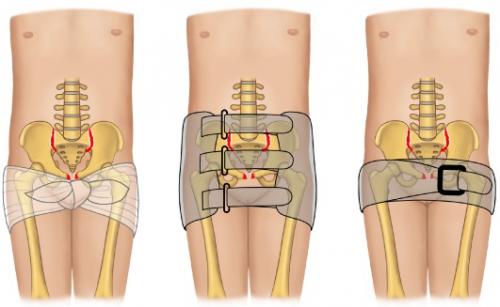
Most patients with suspected pelvic trauma will have a pelvic binder applied pre-hospital. If this is the case, do not remove it unless it is incorrectly positioned or applied. If there is not a correctly-applied binder, then apply one (see below). If in doubt, apply a binder to all major blunt trauma.
During the primary survey, the following should be assessed
- Visual inspection of perineum & genitalia for bruising
- Visual inspection of external urethral meatus, vagina and rectum for bleeding
- Gentle palpation of lower abdomen for tenderness
How and where to apply a pelvic binder
- Make sure you’re familiar with your hospital’s model of binder; the below are general guidelines. If no binder is available, a folded sheet can be used in extremis
- In order to get the binder into the correct position, it can be slid under the knees then worked up to the greater trochanters, or the binder can be placed onto a hospital trolley before transferring the patient from their ambulance trolley
- The binder should be centred over the greater trochanters (see below). The most common mistake is applying it too high, over the ASIS
- As a rule of thumb, tighten the binder until you can just slide a flat hand underneath with moderate pressure

Initial imaging
If a pelvic ring injury is suspected following high-energy trauma, the patient should undergo trauma CT. If the patient is not stable enough for CT, then the binder should remain in place until one can be performed. Plain XR is rarely used now as a first line, but may be used if a patient is too unwell to go through the CT scanner. If a patient has a CT with a binder on, which does not show a pelvic fracture, then they must have one with the binder off as well – sometimes the binder makes fractures invisible, even on CT.
IF THERE IS A CONFIRMED PELVIC RING INJURY, CALL FOR SENIOR HELP IMMEDIATELY
Assessing and treating the pelvis during the secondary survey
The secondary survey should take place after the patient has been initially stabilised as required, and trauma CT has been performed. The primary aim is to assess for evidence of injury to associated structures or open fractures (including bladder/bowel injuries) as well as to identify other injuries.
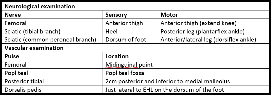
Perform a full lower limb neurological examination:

Assess for an open fracture and/or injury to associated structures:
- Male genitalia: scrotal bruising/haematoma, blood at the meatus
- Female genitalia: labial bruising/haematoma, PV blood, urethral blood. Perform a manual vaginal exam (for lacerations or bony shards)
- Rectum: look for blood and perform a DRE for lacerations or bony shards
See below for guidance on assessment of urological trauma.
Urological trauma & catheterisation
Urological trauma is the most common visceral injury associated with pelvic fractures. There are specific guidelines for its assessment and management as part of the secondary survey.
A single gentle attempt at catheterisation is permissible, even if there is clinical and/or CT evidence of a uretheral injury. This should be performed by an experienced doctor. If you are not happy doing this, liaise with a senior ED, orthopaedic or urological specialist. The guidelines suggest a 16F soft silicone catheter.
If the catheter passes, but drains bloodstained urine, a catheter cystogram should be performed. The details for this are in the guideline.
If the catheter does not pass, then a retrograde urethrogram should be performed. The details for this are in the guideline.
If the catheter passes, but drains frank blood, then do not inflate the balloon. Withdraw the catheter and perform a retrograde urethrogram.
If a catheter cannot be passed, then a suprapubic catheter may be required. Liaise with urology.
Findings of a urine leak from either urethra or bladder suggest an open fracture and the patient should be managed accordingly.
Checklist
- Pelvic binder fitted
- Full primary survey completed
- IV access obtained
- Bloods (FBC, U&E, crossmatch, VBG, clotting) sent and reviewed when back
- IV tranexamic acid given
- Patient resuscitated
- CT obtained
- Secondary survey completed
- Open fracture management initiated if indicated
- Other specialities/teams informed as required (general, vascular, urology, anaesthetics/ICU, theatres)
Admission, discharge and calling a senior
When to call a senior urgently
- Any confirmed pelvic ring injury
- Any suspected pelvic ring injury with haemodynamic compromise, neurovascular deficit, likely open fracture or injury to associated structures
- Any suspected pelvic ring injury where there is a delay to imaging
Who to admit/discharge
- All pelvic ring injuries require admission
- If you are not in an MTC, then liaise with the MTC (after consulting your senior) to determine whether urgent ED-to-ED transfer is indicated
Classification
As mentioned above, the mainstay of imaging is CT. Detailed plain radiograph interpretation is probably not required even of most registrars.
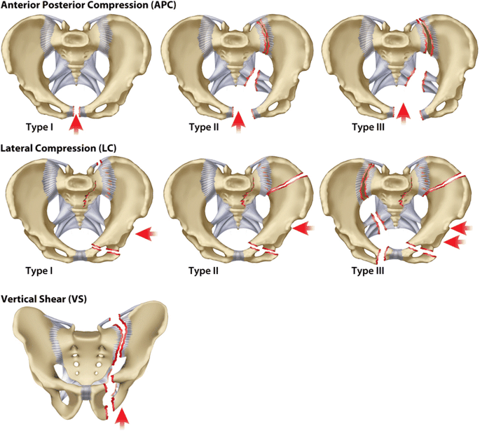
The most commonly utilised classification is the Young &Burgess classification, which is based on the mechanism of injury:

Vertical shear fractures are associated with the highest rates of vascular injury.
Definitive management
The definitive management of pelvic fractures is beyond the scope of this page. Open fractures are managed in the usual way, with early debridement and washout. This may require a defunctioning colostomy.
Temporary stabilisation with an external fixator, or an “in-fix” performing a similar function, may be required until the patient is fit to undergo definitive fixation.
The posterior ring may be reconstructed with screws across the SIJ, or with iliac plating. Anteriorly, a plate may be used across the pubic symphysis.
A period of non-weight-bearing is commonly required.
These patients are high risk for DVT/PE and there should be a plan to prevent, assess for and manage this.
Guidelines
The BOAST for pelvic trauma can be found here, and the BOAST for urological trauma associated with pelvic injuries can be found here.
Page details
Author: Hamish Macdonald
Last updated: 02/04/2020